
Retinal Detachment
One of the most important organs of the human body – the eyes, are prone to develop infections and contract diseases and disorders. Our visionary organ facilitates to see and carry out our daily activities like reading, watching, etc. However, it is important that we take care of our eyes and do not neglect any discomfort that we experience. It is advisable to visit an optometrist and get your checks in case of any abnormalities or irritation.
The eye diseases are generally a result of either a genetic component or some other hormonal or environmental factors.
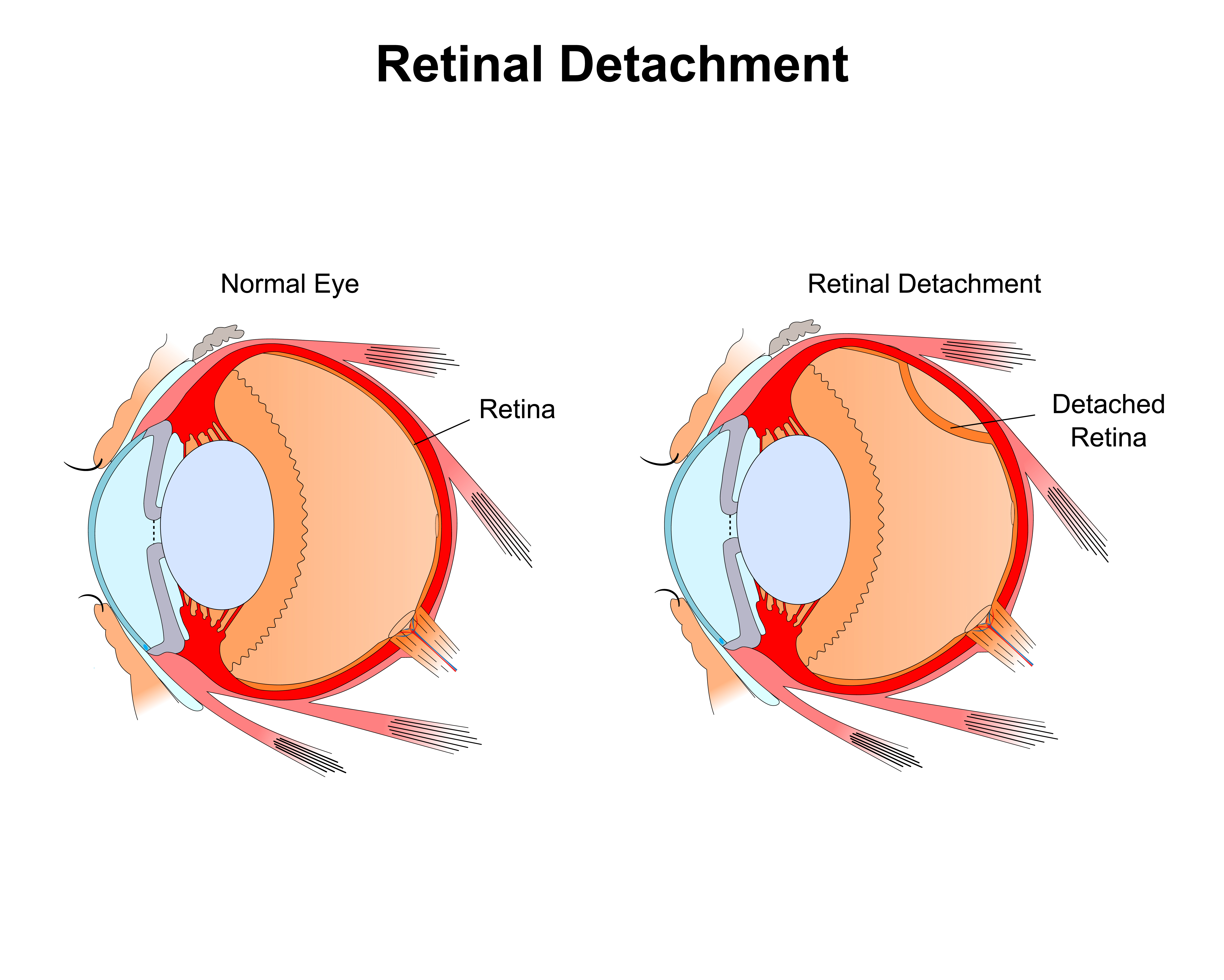
One such eye condition is the retinal detachment. Our eye is made up of extremely delicate, tender and soft components such as the retina. The retina is a covering at the back of the eyeball, which perceives light and generates the electrical signals or impulses to the brain via the optic nerve.
The occurrence of the eye condition is about 5 cases in 100,000 people annually. Retinal detachment is a critical eye condition and the majority of the times it needs surgery to treat it.
What Is Retinal Detachment?
As mentioned previously, the retina in our eye is a thin light-susceptible membrane covering the back line of the eye. The light passing through the lens of the eye focuses the image on our retina which is then responsible for converting the images into signals and generating visual messages that through the optic nerve will be sent to the brain.
Retinal detachment is a condition wherein the retina detaches or separates from the back of the eye and pulls away from its normal position and from the blood vessels responsible for providing oxygen and nourishment. The condition mostly occurs in one of the eyes.
This eye condition is a serious one and should be treated on noticing the signs and symptoms. In case if left untreated for a longer time, the consequences may be permanent eyesight loss.
Causes Of Retinal Detachment
The origins of the emergence of this eye condition may be listed as follows:
An injury to the eye or face can cause retinal detachment.
People with extreme nearsightedness are at the risk of retinal detachment since they have elongated eyeballs with thinner retina.
The vitreous — the gel-like material that fills the inside of the eye- may sag and can lead cause the retina to pull away from its usual position. Also, the fluid movement inside the eye can be a cause of retinal detachment.
People with high diabetes are also susceptible to developing this eye condition.
The emergence of the new blood vessels in the eye can cause the retina to gradually separate from the underlying network of the eye.
Symptoms Of Retinal Detachment
It is to be noted that a person may never experience any pain in case of retinal detachment. However, there are signs of the eye condition developing that are noticeable. These symptoms include:
- Distorted and blurred vision.
- The appearance of floaters, that is, the bits of debris.
- Flashes of light outside the peripheral and central part of the vision. The flashes may appear more in case of increased movement of the eye; This is known as photopsia in medical terms.
- The vision of straight lines appearing curved.
- Heavy eyes.
- The appearance of a shadow from the peripheral vision that slowly and eventually covers the central part of the vision.
- A feeling that a transparent cover is coming down and covering up the entire central vision part of the eye or the appearance of a cloud in the eye.
Types Of Retinal Detachment
The types of retinal detachment can be listed as:
1. Rhegmatogenous Retinal Detachment
It is the most common form of retinal detachment. In this type, there is a hole or a tear in the retina that allows the fluid in the eye that flow to the back of the retina, gradually leading to the retinal detachment.
2. Tractional Retinal Detachment
A scar on the retina surface forces the retina to contract and pull away from the network.
Exudative Retinal Detachment
This type of retinal detachment does not involve any scar or tear, but is developed due to other retinal diseases such as the following:
- A disorder that leads to the fluid in the eye to get to the back of the retina.
- The formation of cancer cells behind the retina.
- The development of an abnormality known as Coats’ diseases that leaks the protein in the blood vessels and build up underneath the retina.
Treatment Of Retinal Detachment
According to a Nashville Optometrist, Dr. Durocher, surgery is required in most of the cases of retinal detachment. Taking the services of an expert Optometrist is advised since the eye condition is a serious one and needs accurate treatment.
The procedures and surgeries performed depend heavily on the type of retinal detachment.
- If a tear or a hole is noticed, but the retina is not detached completely, then photocoagulation with a laser is performed by the optometrist.
- Another method is Cryopexy which involves freezing with intense cold to hold the retina in place.
- Retinopexy is done when slight detachments are observed.
- In case of severe tears and detachments, scleral buckling is performed.
- Vitrectomy is done for larger tears in the retina.
- The scleral buckling is usually performed in combination with vitrectomy.
Related Article: Diabetic Retinopathy Causes Symptoms Prevention And Cure
Retinal detachments usually affect people with age above 40 and people who have diabetes. It is necessary to take care of the symptoms and visit the optometrist for treatment if any signs of retinal detachment are observed.